PhD Project Outline
In October, 2009 I started my PhD in the Molecular Biotechnology group of Prof. Gilles van Wezel at Leiden University. For more information about the research interests of the group, please visit the website: http://science.leidenuniv.nl/index.php/ibl/mbt
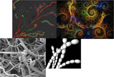
My PhD project involved studying the growth and development of Streptomyces bacteria from both a fundamental and applied perspective, combining molecular biology techniques, light and electron microscopy and computational modeling.
Valuable producers of antibiotics and other bioactive molecules such as enzymes, these multicellular soil-dwelling bacteria are a fascinating topic of study. Streptomyces grow as a branched mycelium and in liquid cultures take on a variety of morphological shapes, from dense pellets to small fragmented mycelia.
In fact, morphology is a determining factor for productivity during industrial fermentations of filamentous organisms, with larger clumps favoring antibiotic production in Saccharopolyspora erythraea, and fragmented clumps stimulating enzyme production by Streptomyces lividans. The understanding and control of mycelial morphology is therefore key for optimization of industrial fermentations.
One way of better understanding morphogenesis is to study the cytoskeleton, or cellular scaffolding, which helps to maintain cell shape, but also plays an important role in both intracellular transport and cell division. One of my research lines therefore involves the study of several putative cytoskeletal proteins and of the effect of their deletion on growth, cell division and morphology.
In collaboration with Prof. Bram Koster from the Leiden University Medical Center (LUMC) and working closely with Dr. Roman Koning, my second research line involved the use of (correlative) cryo-electron tomography to visualize Streptomyces ultrastructure. In this way, we hope to increase fundamental understanding of cell growth, division and morphogenesis, and gain insight into the multicellular lifestyle of Streptomyces. Slices from an initial tomogram, showing complex membranes within a hyphal fragment, are shown below.
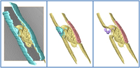
Cryo electron tomograms show that complicated internal structures exist in hyphae.
On the left a tomographic slice through a Streptomyces hyphae is shown, and on the right the internal membranes in the hyphae are surface rendered. Blue is the cell wall, yellow is the membrane, red denotes a flat membrane structure, and small vesicles are shown in purple.
Finally, combining the information gained in the lab and at the microscope, I created a computational model of Streptomyces pellet development.
Such a model may lead to new insights and a better understanding of the relationship between morphology—dictated by microbial growth, branching, fragmentation and adhesion—and product formation. To this end, by combining different modeling methods, I created a structured, 3D morphological modeling framework for rational strain design of Streptomyces species. 2D projections of a pellet simulated using my model are shown below.
The model can be extended based on information gained in fermentation trials for different production strains with the aim to provide a test drive for the fermentation process and to pre-assess the effect of different variables on productivity.
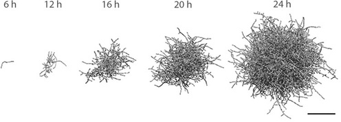
Two-dimensional projection of a 3D simulated pellet. Growth time indicated. Scale bar, 100 μm.
